Have you ever wondered how a biochemistry analyzer “sees” the chemical reaction inside a cuvette?
In this series, we will introduce three common optical systems used in automated biochemistry analyzers.
Today, let’s start with Principle ① — the Filter Wheel Optical System.
A biochemistry analyzer measures biochemical reactions mainly through spectrophotometry.
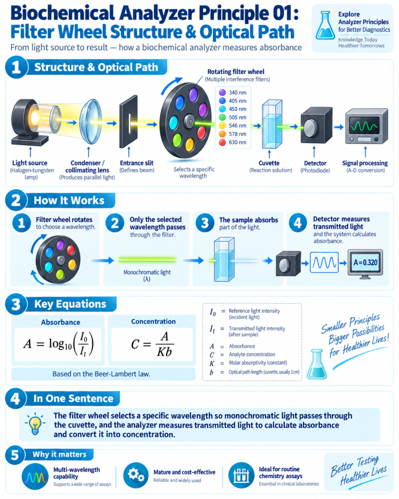
Light from a tungsten-halogen lamp passes through the optical system, and a rotating filter wheel selects the required wavelength, such as 340 nm, 405 nm, 450 nm, 505 nm, 546 nm, 578 nm or 630 nm.
The selected monochromatic light then passes through the reaction solution in the cuvette.
Part of the light is absorbed by the reaction product, while the remaining light reaches the photodetector.
The analyzer compares the incident/reference light intensity I₀ with the transmitted light intensity Iₜ:
A = log₁₀(I₀ / Iₜ)
This relationship is based on the Beer–Lambert Law.
The measured absorbance, or the change in absorbance over time, is then used to calculate the concentration or enzyme activity according to the specific assay method.
Optical Path:
Light Source → Lens/Slit → Filter Wheel → Cuvette → Detector → Signal Processing → Result
The main advantage of the filter-wheel design is its simple structure, reliable wavelength selection and relatively low cost, making it widely used in routine clinical chemistry analyzers.
In the next post, we will look at Biochemistry Analyzer Principle ② — another wavelength separation method.
#BiochemistryAnalyzer #ClinicalChemistry #LaboratoryEquipment #IVD #Spectrophotometry #MedicalLaboratory #LabTechnology